Recurrent low back pain in a young athlete
3 Tesla MRI in the assessment of overload-related low back pain
Episodic low back pain in young athletes is often related to functional load, but when small disc bulges or bony remodeling are seen for the first time, they can lead to overly alarming interpretations. In this clinical case, a preventive MRI performed two years earlier proved essential to understand the overall picture and to guide clinical management.
The case: a young athlete with low back pain during a high-load phase
The patient is a competitive athlete who, two years earlier, had undergone a check-up lumbar MRI in the complete absence of symptoms. The scan showed small disc bulges and mild signs of inflammatory-type bony remodeling involving the vertebral endplates: findings that are not common at that age, but compatible with intense sports activity.
During a new period of very high training load, the athlete developed bilateral low back pain, with discomfort radiating to the left gluteal region. Symptoms were not accompanied by neurological signs, but they interfered with training.

Preventive lumbar MRI: small disc bulges and inflammatory-type bony remodeling, stable and not clinically significant.
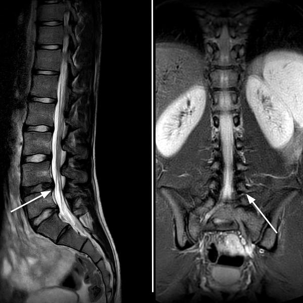
MRI during pain: similar morphological appearance, no new herniation, and no signs of spondylolisthesis or instability.
What does the MRI show?
A direct comparison between the two MRIs demonstrates complete stability of the findings: no additional herniation, no progression of the disc bulges, and no signal suggesting acute edema or vertebral instability. This allowed the pain to be correctly interpreted as a load-related functional episode, rather than a new structural injury.
Why does this happen?
In young athletes exposed to intense workloads—especially in explosive disciplines—the lumbar spine is subject to pressure changes and traction forces that can generate symptoms without creating true lesions. Small disc bulges that remain stable over time often represent a functional adaptation, not a progressive disease.
Athlete management
The clinical and radiological picture supported a conservative approach:
- No need to completely stop sports activity
- A short course of a mild anti-inflammatory medication
- Temporary modulation of training loads
- Gradual return to activity once pain-free
Conclusions
This case shows how a preventive MRI performed in a symptom-free phase can become an invaluable reference. Without that prior scan, the findings observed during the painful episode would likely have generated clinical anxiety and suspicion of an acute disc injury, leading to prolonged rest or unnecessary treatments.
3 Tesla MRI proves to be a key tool not only for detecting pathology, but also for identifying what is not pathological, guiding safer and more rational management in young competitive athletes.